After our earlier consideration of studies aimed at detecting point mutations and clastogens in parts 1 and 2 of this series, we come onto the last broad class of mutagens – aneugens. As mentioned, mutagenic substances can result in inheritable diseases, cancer, metabolic diseases and ageing, so they are important in safety testing.
Aneugens are substances that affect a specific stage of the cell cycle, leading to a change in the number of chromosomes in a cell, typically due to interaction with cellular machinery involved in separating chromatids during anaphase. This is a mechanistically distinct class from mutagenic compounds via direct DNA base interaction, interference in DNA replication or chromosome structure. Aneuploidy can happen spontaneously in cells and might also be part of mechanisms of other DNA-altering events, such as chromothripsis. However, several classes of well-known aneugenic chemicals are potentially risky to a man if exposure is high enough, resulting in genetic diseases in offspring, embryo lethality or cancer.
Because aneugens have carcinogenic and reproductive toxicity potential, spotting agents with these properties early during the development of new substances is of high importance. While it is generally accepted that aneugens have thresholds (because it usually takes multiple hits to the cell spindle to result in non-disjunction), they can be potent substances and be challenging to risk assess.
The significance of aneugenicity depends on the industry sector.
For high-volume chemicals administered under REACH, aneugenic effects leading to reproductive or carcinogenic outcomes may be subject to more restricted uses or containment procedures or uses may be prohibited. In other areas, such as pharmaceuticals, aneugenic properties may be considered acceptable for high-risk patients but could result in the drug not being licensed for patients at lower risk or of reproductive age.
Developing a new pesticide to generate a global data package will often include developmental and multi-generational toxicity studies conducted to high doses for hazard assessment. Findings of compound-related embryo lethality or birth defects could result in a hazard classification that leads to an outright ban on active use in regions such as Europe – even if the effects are confined to high doses and the substance otherwise passes a risk assessment. As such, an earlier assessment of the potential for a substance to induce aneuploidy is of relevance from not just a mutagenicity perspective.
Predicting aneugenicity without animal models
As in vivo developmental and reproductive toxicity studies and chronic toxicity studies (plus the range-finding studies that need to conduct them) use large numbers of animals, are time-consuming and use kilograms of test items, we need other ways of identifying and ideally screening out aneugens.
Some aneugenic substances can be flagged by structure-activity (SAR) in silico predictions due to structural similarity to known examples, but typically they need to be detected in vitro genotoxicity studies to reliably spot them. In some cases, and where it’s permissible to do so, in vivo studies can be conducted as a follow-up to see if substances positive in vitro express this activity in vivo, and if they do, can be used to determine a no-effect level that can help inform a risk assessment. Understanding the mechanistic link between an in vitro genotoxicity positive and an aneugenic mechanism is key if such an approach is to be utilised.
The in vitro test primarily used for detecting aneugenicity is the in vitro micronucleus assay. Micronuclei are formed from chromosomes or fragments of chromosomes that have not properly been incorporated into the nucleus during cell division. These whole chromosomes or fragments become membrane-bound and may remain within the daughter cells after the point of cytokinesis during cell division. When visualised under a microscope, they appear as micronuclei adjacent to the main nucleus. A range of rodent and human cell lines, as well as primary cells, are accepted for use in the performance of this test. At Gentronix, we conduct GLP OECD guideline 487 in vitro micronucleus studies in TK6 cells or human lymphocytes to meet our clients’ regulatory needs.
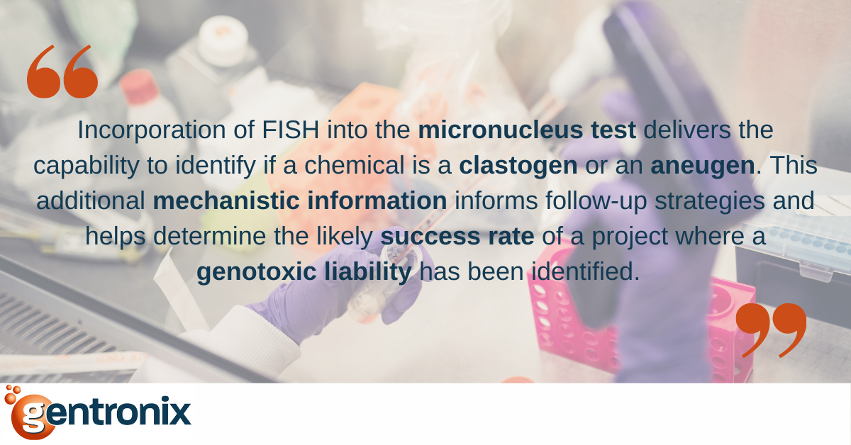
As discussed in Part 2 of this series, the micronucleus assay can detect clastogens (substances causing structural but not numerical changes to chromosomal structures) and aneugens. To differentiate these two mechanisms, the presence or absence of centromeres or kinetochores within micronuclei can be assessed using methods such as FISH staining (fluorescence in situ hybridisation) with pan-centromeric probes or CREST anti-kinetochore labelling. If centromeres or kinetochores are present, the observed micronucleus is more likely to contain a whole chromosome (evidence of aneugenicity); without it, it indicates a micronucleus formed from a chromosome fragment (as a result of clastogenicity). Within regulatory testing, typically a hundred micronuclei are assessed for the presence or absence of centromeres/kinetochores per dose, with aneugenic substances inducing typically >50 – 95% centromere/kinetochore containing micronuclei.
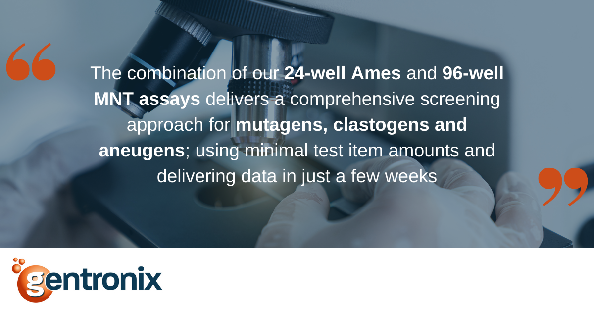
Detection of an aneugenic mechanism is also possible in higher throughput screening formats. Tools such as MultiFlow™, which uses multiple endpoints to differentiate clastogens from aneugens, allow the detection of genotoxicity and differentiation of mode of action (MoA) in the same test. By introducing a miniaturised 96-well format of the in vitro micronucleus assay, we can conduct studies in the same manner as a GLP OECD guideline study but greatly save on test item use. This miniaturised assay is compatible with FISH staining, enabling aneugens to be differentiated from clastogens at the compound screening stage. Both of these services to establish MoA for clastogenic and aneugenic materials are offered by Gentronix.
Some authors have published evidence that the L51 (mouse lymphoma assay) can detect aneugens as well as point mutations and clastogenicity, but it is generally not recommended for this purpose. Other commonly used genotoxicity assays, such as the Ames or Comet assays, will not detect aneugens, and this lack of mechanistic coverage should be considered when designing testing strategies.
Whilst not specifically detecting aneugens, the GreenScreen HCTM and BlueScreen HC® GADD 45-alpha assays have been demonstrated to have very high sensitivity (i.e. high positive predictivity) for genotoxins, including aneugens, whilst having high specificity (high negative predictivity), and are excellent options for early-stage screening as they can be conducted with just 10 mg of the test item. Being gene reporter assays, GreenScreen and BlueScreen measure the cell’s response to DNA damage rather than mutation per se. They provide a high-throughput screening option, which can be used as a triage tool, enabling follow-up in an in vitro micronucleus test with FISH for positive results suspected of being driven by an aneugenic mechanism.
In vivo tests for detecting aneugens
Consideration of potential aneugenicity of a compound is not restricted to in vitro testing, and where regulatory environments allow or mandate, the in vivo test typically used to assess aneugenicity is the in vivo bone marrow micronucleus assay (OECD guideline 474). This assay looks at the potential for a test substance to induce micronuclei within the haematopoietic system, with both bone marrow and peripheral blood able to be analysed for the presence of micronuclei. This indicates the potential systemic clastogenic and aneugenic risk of a substance within the animal and, therefore, the potential risk to human health. FISH/anti-kinetochore labelling can also be used in vivo studies to differentiate clastogens and aneugens. However, this is currently restricted to microscopy-based methods focusing on the bone marrow compartment. Later in 2019, Gentronix will add the rat bone marrow micronucleus assay to its service list, with slide scoring of bone marrow smears or flow cytometric peripheral blood analysis available. Additionally, we will partner with other expert companies to provide the bioanalysis that can demonstrate test item is systemically available using blood or plasma analysis as required.
In vivo bone marrow micronucleus test data can be utilised alongside in vitro data to demonstrate the potential safety of a substance where aneugenicity has been confirmed as the overriding mechanism of the toxicological event. Statistical analysis of the in vivo data dose-response profiles can be used to establish Benchmark Dose profiles. Establishing acceptable safety margins is possible when the exposure and/or efficacy data allow.
Other commonly used in vivo assays, such as the Comet and transgenic assays, are unsuitable for assessing aneugenicity, as they provide mechanistic coverage for mutation induced by DNA reactivity &/or clastogenicity. When conducting an in vivo assay, it is essential to consider whether there is evidence that the tissue being assessed is adequately exposed to the test substance or its relevant metabolites for the results to be meaningful.
When performing genotoxicity safety assessments, it is important to consider the detection of all mutagenic mechanisms, including aneugenicity. Over the last ten years, regulatory approaches across industries have sought to include the assessment of potential aneugenicity into the required genotoxicity testing approaches through the micronucleus test. Whilst having the primary aim of further protecting human health and the environment, this also provides the option for management of adverse findings, with aneugenic mechanisms accepted as having thresholds of no effect, and therefore enabling potential mitigation of positive genotoxicity findings by confirmation of this mechanism and establishment of a no-observed-genotoxic-effect-level dose (NOGEL) increasingly being possible. However, the challenge remains to extrapolate aneugenic no-effect levels to scenarios of early life stages and over periods of longer-term exposure (such as developmental and multi-generational studies). The tools for detecting and assessing aneugens have come a long way, and with tools such as FISH and MultiFlowTM, they are continuing to get better and easier to perform.