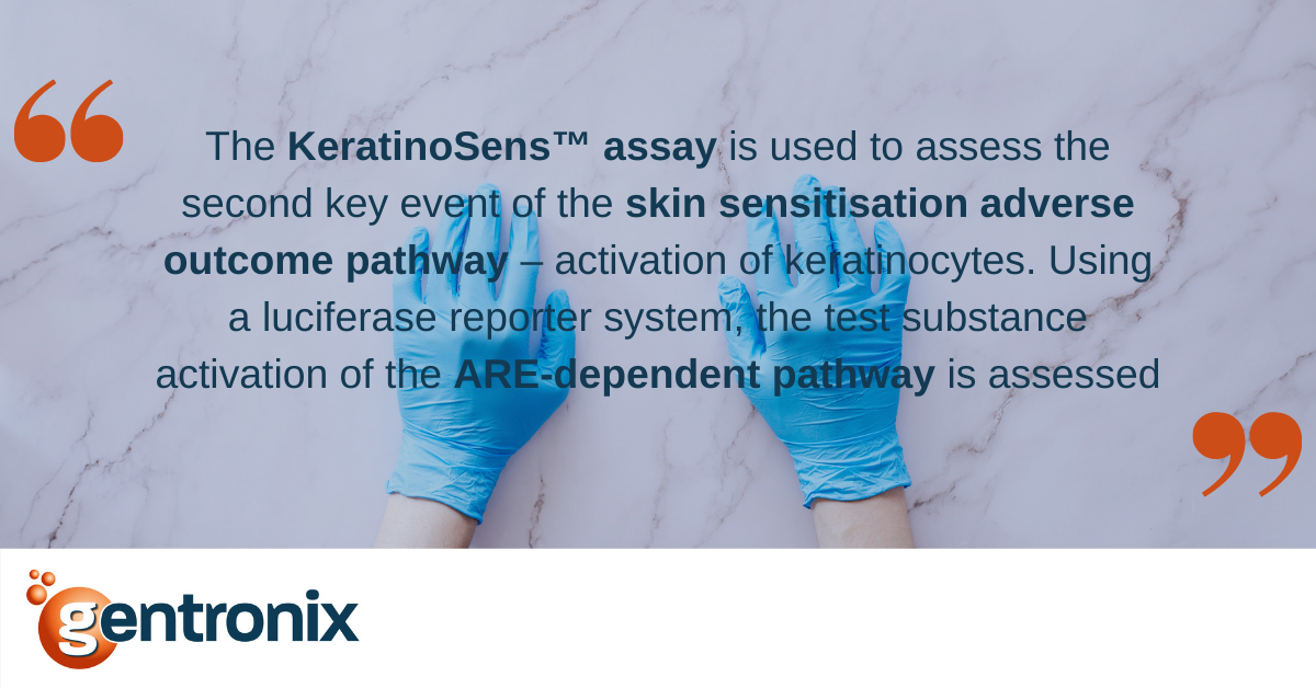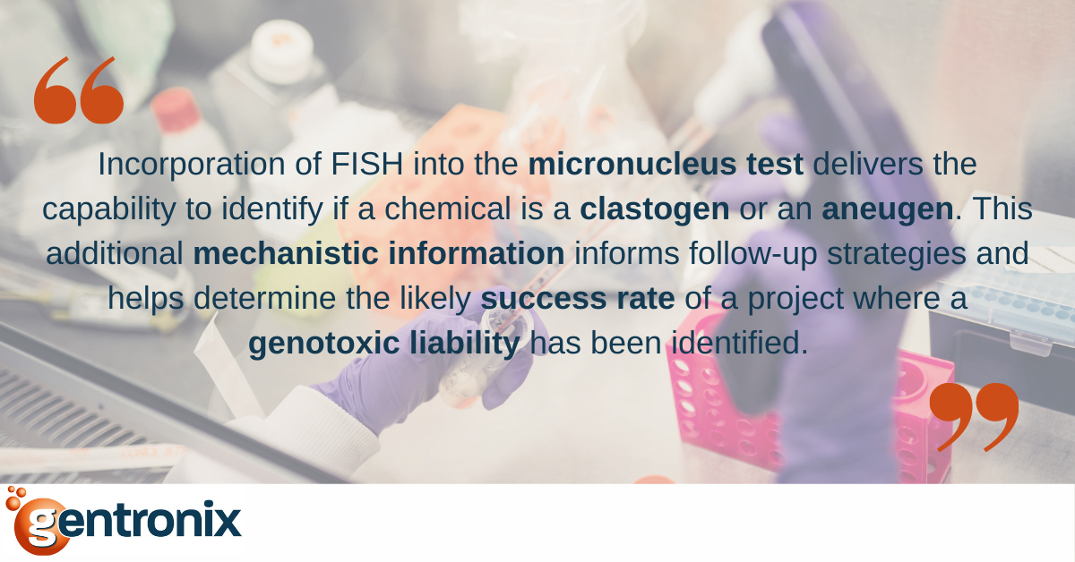
In addition to assessing a chemical’s ability to induce mutations, typically using the bacterial Ames test, a key aspect of understanding risk to human health is assessing the potential genotoxicity within mammalian cells. These assessments, especially where an Ames test has returned a negative result, are designed to assess the liability of a chemical to induce chromosomal damage in the form of either break (clastogens) or missegregation and whole chromosome loss (aneugens). Such assessments are commonly conducted using the in vitro and in vivo micronucleus tests, as these tests have high sensitivity for genotoxic substances and can detect both clastogens (direct and indirect mechanisms) and aneugens. Importantly, these studies can also be adapted to determine whether a positive result results from clastogenic or aneugenic modes of action.
Where test substances cause genotoxic stress through interference within mitosis, either through tubulin stabilisation/destabilisation or interference in mitotic kinase cascades, the result is often a missegregated whole chromosome that becomes a micronucleus. A distinct feature of this lagged chromosome is that it contains centromeric sequences within the DNA sequence and the presence of the kinetochore protein assembly. Where a substance induces dsDNA breaks (clastogens), the resulting micronuclei will be a mix of chromosomal fragments, many of which will lack the centromeric sequences and kinetochore protein complex. This distinction in chromosome structure within a micronucleus can be exploited to determine if the majority of micronuclei are centric and indicative of an aneugenic mode of action or acentric and a result of clastogenic mechanisms.
Within the OECD 487 in vitro micronucleus test using human-derived TK6 cells, the centromeric regions can be probed using fluorescence in situ hybridization (FISH) techniques that hybridise across the centromeric regions of all human chromosomes. With DNA labelled using a counterstain such as DAPI, nuclei and micronuclei can be enumerated using fluorescence microscopy, and the presence or absence of FISH centromere labelled micronuclei can be determined. Where centric micronuclei are >65% of all micronuclei analysed, the likelihood is that a genotoxic substance inducing micronuclei is predominate via an aneugenic MoA.
The same techniques to distinguish clastogens and aneugens can be incorporated into the OECD 474 in vivo micronucleus test, though at present, FISH techniques are restricted to mouse studies. Within the rat, whole chromosomes are identified using antibodies raised to the kinetochore assembly using CREST techniques. This ability to detect both genotoxic stresses, identify their mode-of-action in vitro through the incorporation of FISH, and then determine if the mode-of-action translates to the in vivo setting is a powerful advantage of the micronucleus test. It assesses the potential genotoxicity of test substances and their relevance to in vivo and human health settings.